Click images to enlarge.
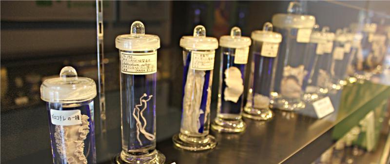
Laika ac CC-BY-SA 2.0


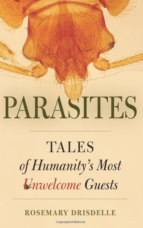


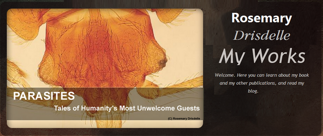
Drisdelle
has written one
of those rare books that is fun to read but does not skimp on scholarly rigor...
(Janice Moore)
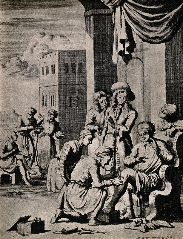
A surgeon extracts a Guinea worm from a man's leg (another patient and extracted worm in the background). Engraving by Jan Luiken (1649 - 1712); Wellcome Images V0016741 CC BY 4.0